1.2 Elements of Neuronal Dynamics
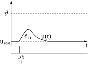
The effect of a spike on the postsynaptic neuron can be recorded with an intracellular electrode which measures the potential difference between the interior of the cell and its surroundings. This potential difference is called the membrane potential. Without any input, the neuron is at rest corresponding to a constant membrane potential . After the arrival of a spike, the potential changes and finally decays back to the resting potential, cf. Fig. 1.5A. If the change is positive, the synapse is said to be excitatory. If the change is negative, the synapse is inhibitory.
At rest, the cell membrane has already a strongly negative polarization of about –65 mV. An input at an excitatory synapse reduces the negative polarization of the membrane and is therefore called depolarizing. An input that increases the negative polarization of the membrane even further is called hyperpolarizing.
1.2.1 Postsynaptic Potentials
Let us formalize the above observation. We study the time course of the membrane potential of neuron . Before the input spike has arrived, we have . At the presynaptic neuron fires its spike. For , we see at the electrode a response of neuron
| (1.1) |
The right-hand side of Eq. (1.1) defines the postsynaptic potential (PSP). If the voltage difference is positive (negative) we have an excitatory (inhibitory) postsynaptic potential or short EPSP (IPSP). In Fig. 1.5A we have sketched the EPSP caused by the arrival of a spike from neuron at an excitatory synapse of neuron .
| A | ||
|---|---|---|

|
|
|
| B | ||

|
|
|
| C | ||

|
 |
1.2.2 Firing Threshold and Action Potential
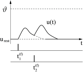
Consider two presynaptic neurons , which both send spikes to the postsynaptic neuron . Neuron fires spikes at , similarly neuron fires at . Each spike evokes a postsynaptic potential or , respectively. As long as there are only few input spikes, the total change of the potential is approximately the sum of the individual PSPs,
| (1.2) |
i.e., the membrane potential responds linearly to input spikes; see Fig. 1.5B.
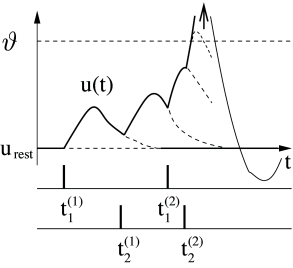
On the other hand, linearity breaks down if too many input spikes arrive during a short interval. As soon as the membrane potential reaches a critical value , its trajectory shows a behavior that is quite different from a simple summation of PSPs: The membrane potential exhibits a pulse-like excursion with an amplitude of about 100 mV. This short voltage pulse will propagate along the axon of neuron to the synapses with other neurons. After the pulse the membrane potential does not directly return to the resting potential, but passes, for many neuron types, through a phase of hyperpolarization below the resting value. This hyperpolarization is called ‘spike-afterpotential’.
Single EPSPs have amplitudes in the range of one millivolt. The critical value for spike initiation is about 20 to 30 mV above the resting potential. In most neurons, four spikes – as shown schematically in Fig. 1.5C – are thus not sufficient to trigger an action potential. Instead, about 20-50 presynaptic spikes have to arrive within a short time window to trigger a postsynaptic action potential.